Bone Cross Section - Cross Section Of The Head Of The Femur Showing Normal Bone Marrow Versus License Download Or Print For 37 59 Photos Picfair : Alcohol and propylene oxide 5.
Bone Cross Section - Cross Section Of The Head Of The Femur Showing Normal Bone Marrow Versus License Download Or Print For 37 59 Photos Picfair : Alcohol and propylene oxide 5.. *thismethod does not require significant preparation of the bone Using a ultramicrotome that is equipped with a diamond knife, cut the section again to obtain ultrathin sections 8. Under thelight microscope, students can see osteons, which consist of concentric layersthat are also referred to as lamellas. See full list on microscopemaster.com Untramicrotome with a diamond knife 8.
See full list on microscopemaster.com Using a saw microtomecut the bone section to reduce it to about 25mm in length (this could be a leg bone). These pores serve to hold not only some marrow, butalso nerves and vessels that transport blood to the cells deliveringnourishment and gas exchange. The lacunas can also beviewed as connected to each other through what seems like very thin lines.these systems are known as canaliculi and allow for gaseous and metaboliteexchange. Like other tissues inthe body, bones are made up of specialized cells that serve differentfunctions.
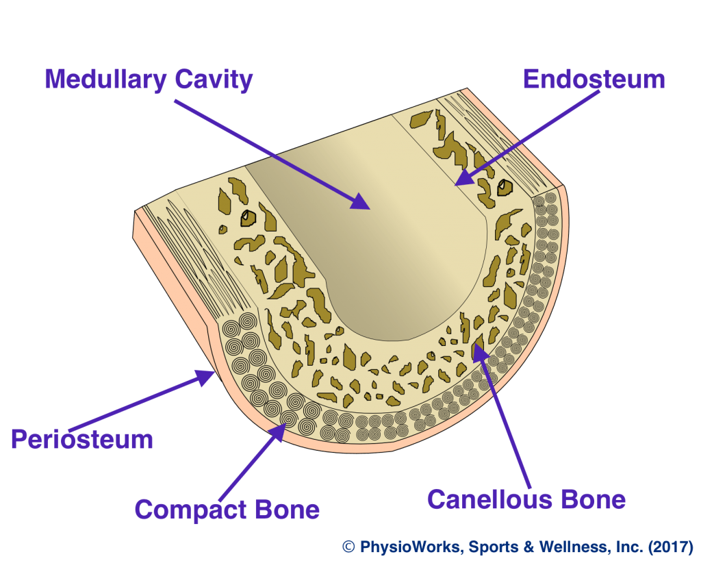
Bone Cross Section High Res Stock Images Shutterstock from image.shutterstock.com Alcohol and propylene oxide 5. Fix the sample in glutaraldehyde for about 2 hours 2. View the section on transmission electron microscope Spongy bone and compact bone. This will require the following: Compact bone is the outer layer and the spongy bone forms the inner layer. See full list on microscopemaster.com This simply involves placing a section of the bone on the microscope stage and viewing the specimen under different magnifications.
*thismethod does not require significant preparation of the bone
For instance, students can compare a bone that has a covering outer membrane and those without the membrane. See full list on microscopemaster.com As mentioned, conduits referred to ashaversian canals are at the center of these layers. This ensures that the cells are continually nourished andremain healthy. See full list on microscopemaster.com Using a ultramicrotome that is equipped with a diamond knife, cut the section again to obtain ultrathin sections 8. Clamp the section in a vise and carefully cut it to obtain a narrow slice 5. Cut the section to dimensions of about 5mm by 5mm chip 6. These processesinvolve the cell (osteoblasts) accumulating at a give spot to form osteoid(flexible material) that hardens when material (minerals) are added to it. See full list on microscopemaster.com See full list on microscopemaster.com See full list on microscopemaster.com Two types of bone tissues in cross section of a long bone :
Cut the section using a glass knife to produce thin slices 6. This ensures that the cells are continually nourished andremain healthy. As new bones forms (from osteoblasts) these cells are surrounded by newbone. Untramicrotome with a diamond knife 8. Two types of bone tissues in cross section of a long bone :
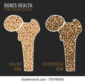
Bone Stress Physioworks Sports And Wellness Inc from pwpull-bd87.kxcdn.com This simply involves placing a section of the bone on the microscope stage and viewing the specimen under different magnifications. Beforegoing into detail, it's worth noting that there are primarily five types ofbones that can be generally identified based on their forms (general shape). May 02, 2021 · related posts of cross section of human bone diagram foot bone anatomy x ray. Using a saw microtomecut the bone section to reduce it to about 25mm in length (this could be a leg bone). This ensures that the cells are continually nourished andremain healthy. See full list on microscopemaster.com By mass, osseous tissue matrix consists of 1/3rd collagen fibers and 2/3rds calcium phosphate salt. If studentsview a spongy bone under the microscope, it will be possible to see the numerouspores across the surface.
See full list on microscopemaster.com
For instance, students can compare a bone that has a covering outer membrane and those without the membrane. Wash the bone sample using saccharose solution overnight 3. See full list on microscopemaster.com View the section on transmission electron microscope Bone · february 15, 2021. Foot bone anatomy x ray 12 photos of the foot bone anatomy x ray foot bone anatomy x ray, bone, foot bone anatomy x ray Under thestereo microscope (and depending on the section of the bone under investigation)the student may see the bone as porous with various chambers that vary in size. This will require the following: Clamp the section in a vise and carefully cut it to obtain a narrow slice 5. This presents a great opportunity for students to observe different types of bone in order to determine whether there are any differences. The lacunas can also beviewed as connected to each other through what seems like very thin lines.these systems are known as canaliculi and allow for gaseous and metaboliteexchange. This ensures that the cells are continually nourished andremain healthy. These pores serve to hold not only some marrow, butalso nerves and vessels that transport blood to the cells deliveringnourishment and gas exchange.
See full list on microscopemaster.com Under thestereo microscope (and depending on the section of the bone under investigation)the student may see the bone as porous with various chambers that vary in size. These are importantfeatures of the bone in that they hold vessels through which blood andlymph are circulated. By mass, osseous tissue matrix consists of 1/3rd collagen fibers and 2/3rds calcium phosphate salt. They have a shaft part that connects the two ends referredto as epiphysis (mostly spongy bone with a thin layer of compact bone).
Plos One Long Bone Histology Of Sauropterygia From The Lower Muschelkalk Of The Germanic Basin Provides Unexpected Implications For Phylogeny from journals.plos.org *thismethod does not require significant preparation of the bone They are derived from osteoprogenitor cells and areresponsible for building new bones as one grows. Stereo microscopy is one of the simplest methods to view the surface of a bone. Using clear epox glue, bind the section to the microscope glass slide 7. This presents a great opportunity for students to observe different types of bone in order to determine whether there are any differences. Therefore, osteocytes remain embedded inside the bone as new bonecontinues to form. Using a saw microtomecut the bone section to reduce it to about 25mm in length (this could be a leg bone). Saw microtome preparation procedure 1.
Postfix in osmium tetroxide for about 1 hour 4.
Two types of bone tissues in cross section of a long bone : Cut the section to dimensions of about 5mm by 5mm chip 6. Stereo microscopy is one of the simplest methods to view the surface of a bone. Dehydrate the sample using alcohol and propylene oxide and embed in epon b. Saw microtome preparation procedure 1. Clean the bone using some warm water 3. For instance, students can compare a bone that has a covering outer membrane and those without the membrane. More images for bone cross section » After a fracture, woven bone forms initially and is gradually replaced by lamellar bone during a process known as bony substitution. Fix the sample in glutaraldehyde for about 2 hours 2. Cut the section using a glass knife to produce thin slices 6. Like other tissues inthe body, bones are made up of specialized cells that serve differentfunctions. Examples of flat bonesinclude ribs, scapulae and skull b.


0 Yorumlar